
Cell Structure and Function
Cell theory, prokaryotic and eukaryotic cells, why cells are small, unicellular and multicellular life, cell specialization and the origin of organelles.
🎯 By the end of this lesson
- State the three parts of cell theory.
- Compare light and electron microscopes in terms of magnification, resolution and use with living cells.
- Calculate magnification or actual size from a drawing or micrograph.
- Compare prokaryotic and eukaryotic cells using structures present, size and examples.
- Identify the function of the major organelles and describe how they cooperate to make and export a protein.
- Explain, using surface area-to-volume ratio, why cells are small.
- Explain how cells of one organism can have the same DNA but different structures and jobs.
- Summarize the evidence for the endosymbiotic origin of mitochondria and chloroplasts.
1Overview
Every living thing on Earth, from the bacterium on a doorknob to the blue whale, is built from cells. A whale cell and a bacterial cell, though, differ as much as a city differs from a one-room workshop. Why did life settle on the cell as its building block? What separates the two great cell plans? And how can one fertilized egg turn into hundreds of different kinds of cells that all carry the same instructions?
2Cell theory and the tools that revealed cells
Cells were unknown until microscopes improved enough to show them. By the nineteenth century, Schleiden, Schwann and later Virchow had combined many observations into the cell theory, which has three main statements:
- All living things are made of one or more cells.
- The cell is the basic unit of structure and function in life.
- New cells arise only from existing cells.
Two features of microscopes matter for reading cell diagrams. Magnification is how much larger the image appears than the real object. Resolution is the ability to tell two nearby structures apart as separate. Magnifying a blurry image only makes a larger blur; a better resolution is what reveals new detail.
| Feature | Light microscope | Electron microscope |
|---|---|---|
| Illumination | Visible light | Beam of electrons |
| Typical magnification | Roughly 400x in a school lab, about 1000x with oil immersion | Up to about 100,000x |
| Resolution | About 200 nm | Far finer, in the picometre range |
| Living specimens | Can be viewed alive (staining usually kills them) | Specimens are killed |
| Types | Compound, stereo | Scanning (surface) and transmission (interior) |
Magnification = image size ÷ actual size. A drawing of a protist is 60 mm across and the real cell is 0.12 mm wide. Magnification = 60 ÷ 0.12 = 500x. If an actual size is wanted instead, rearrange: actual size = image size ÷ magnification, so a 60 mm image at 500x represents 0.12 mm. Units must match before dividing (1 mm = 1000 µm, so 0.12 mm is 120 µm).
The history of cell biology follows the history of the microscope. Light microscopes revealed cells and the nucleus. Electron microscopes later revealed the internal membranes, ribosomes and the double membranes of mitochondria. Every improvement in the tool changed what biologists could ask.
3Two cell plans: prokaryotic and eukaryotic
All cells have a plasma membrane (the outer boundary), cytoplasm (a gel-like interior), ribosomes (which build proteins) and DNA (the genetic information). Beyond these shared parts, cells divide into two plans.
Prokaryotic cells
Prokaryotes, the Bacteria and Archaea, are mostly single-celled and lack a nucleus and other membrane-bound organelles. Their circular chromosome sits in a central region called the nucleoid, which has no membrane around it. Most bacteria also have a cell wall of peptidoglycan that protects the cell and maintains its shape, and many have a polysaccharide capsule outside the wall that helps them attach to surfaces. Flagella move the cell, fimbriae help attach to host cells, and pili can transfer genetic material to another bacterium by direct contact, a process called conjugation (covered in the next lesson).
Eukaryotic cells
Eukaryotes (animals, plants, fungi and protists) have a nucleus and other membrane-bound organelles, compartments that each specialize in a job. Eukaryotic cells are typically much larger than prokaryotic cells: about 10 to 100 µm across versus about 0.1 to 5 µm. The main organelles are summarized below.
| Structure | Function |
|---|---|
| Nucleus | Holds DNA and directs protein synthesis; enclosed by a double-membrane envelope with pores; the nucleolus assembles ribosome subunits |
| Rough ER | Ribosome-studded; makes membrane and secretory proteins |
| Smooth ER | Makes lipids |
| Golgi apparatus | Stacks of flattened membranes that modify proteins and lipids from the ER |
| Mitochondrion | Cellular respiration; makes ATP; double membrane with inner folds (cristae); has its own DNA and ribosomes |
| Chloroplast (plants, algae) | Photosynthesis; chlorophyll in thylakoids stacked as grana, sugar made in the stroma |
| Lysosome (animal cells) | Digestive enzymes break down worn-out organelles and materials |
| Vacuoles and vesicles | Storage and transport; the plant central vacuole holds water and keeps pressure against the cell wall |
| Cytoskeleton | Microfilaments, intermediate filaments and microtubules; shape, organelle position, the mitotic spindle |
| Cell wall (plants, fungi, some protists) | Protection and support; cellulose in plants |
Plant cells do not have "mitochondria instead of chloroplasts" or the reverse. Plant cells have both. Chloroplasts make sugar from light; mitochondria release the energy of that sugar as ATP. Animal cells have mitochondria only.
| Feature | Prokaryotic | Eukaryotic animal | Eukaryotic plant |
|---|---|---|---|
| Nucleus | No (nucleoid) | Yes | Yes |
| Membrane-bound organelles | No | Yes | Yes |
| Cell wall | Usually (peptidoglycan in bacteria) | No | Yes (cellulose) |
| Chloroplasts | No (some bacteria photosynthesize without them) | No | Yes |
| Lysosomes | No | Yes | Most lack them |
| Typical size | 0.1 to 5 µm | 10 to 100 µm | 10 to 100 µm |
Organelles as a production line
Organelles cooperate like stations in a factory. Following one secreted protein, such as a digestive enzyme, shows the teamwork:
- The nucleus holds the gene, and its DNA is transcribed into RNA that leaves through a nuclear pore.
- Ribosomes on the rough ER translate the RNA into protein, which enters the ER.
- Vesicles carry the protein to the Golgi apparatus, which modifies it.
- Further vesicles carry the finished product to the plasma membrane or to a lysosome.
- Mitochondria supply ATP for all of these steps, and the cytoskeleton provides tracks and shape throughout.
The loss of any station disrupts the whole line. This is why a mutation affecting a single organelle protein can cause disease at the level of tissues and organs.
Compartments allow a cell to run many reactions at once without interference. Lysosomes keep powerful digestive enzymes in a more acidic compartment so that they do not digest the rest of the cell. Prokaryotes get by without compartments mainly because their cells are tiny.
4Why cells are small: surface area to volume
Why are cells microscopic? A cell must take in nutrients and release wastes across its surface, but the cell's needs depend on its whole volume. As a cell grows, volume rises faster than surface area, so the surface area-to-volume ratio falls. Beyond a certain size the membrane cannot supply the interior fast enough. Small cells allow ions, nutrients and wastes to diffuse quickly throughout the cell.
Cells cope in several ways: they stay small, divide, fold their membranes (the cristae of a mitochondrion, for example), become thin or elongated, or develop internal compartments. Organelles are one reason eukaryotic cells can be larger than prokaryotic cells.
A cube-shaped cell has sides of 2 units. Surface area = 6 × 22 = 24. Volume = 23 = 8. Ratio = 24 ÷ 8 = 3. If the sides triple to 6 units, surface area = 6 × 36 = 216 and volume = 216, giving a ratio of 1. The bigger cell has only one third of the surface per unit of volume, so diffusion alone would supply it much less well.
5Unicellular and multicellular organisms
A unicellular organism is a single cell that does everything: it takes in energy, grows, responds to its environment and reproduces. Bacteria, archaea, many protists and yeasts are unicellular. A multicellular organism is built of many cells that cooperate. Because the whole body does not need to be bathed directly in the environment, a multicellular body can grow large while each cell stays small enough to keep a workable surface area-to-volume ratio.
Prokaryotes are the most abundant life in every habitat and include extremophiles living in hot springs, deep ocean, salt lakes, polar ice and acidic sites. Unicellular life is not "simple": it was the only life on Earth for billions of years, and a single prokaryote can do chemistry that no animal can.
Humans carry far more prokaryotic cells than many people expect: one textbook estimate is that bacterial cells in the body outnumber human cells roughly ten to one. See the article on the human microbiome.
Advantages and costs of each body plan
| Body plan | Advantages | Limits |
|---|---|---|
| Unicellular | Large surface area relative to volume; rapid exchange with the surroundings; can live almost anywhere | Size is limited; each cell must perform every function; usually exposed directly to the environment |
| Multicellular | Can grow large; division of labour among specialized cells; internal environment can be regulated | Needs transport and communication systems; cells depend on one another; damage to key tissues can be fatal |
Cooperation among prokaryotes: biofilms
The line between unicellular and multicellular is not always sharp. Many prokaryotes form biofilms, communities held together by a matrix made mostly of polysaccharides. Biofilm cells attach to surfaces, such as the inside of a pipe, and compared with free-floating cells they resist antibiotics and disinfectants better. A biofilm is still a community of independent cells rather than a body with tissues, but it shows how cooperation can offer a survival advantage, which is one of the ideas behind the evolution of multicellularity.
Question: A cell has a nucleus, a cell wall made of cellulose, chloroplasts and a large central vacuole. Name its kingdom-level type and explain how it differs from a bacterium.
Answer: It is a plant cell (eukaryotic). Unlike a bacterium it has a membrane-bound nucleus and organelles, and its wall is made of cellulose and not peptidoglycan. It is also likely 10 to 100 µm across, much larger than the typical bacterial cell.
6Cell specialization in multicellular organisms
A body contains hundreds of cell types: neurons, red blood cells, muscle cells, skin cells, root hair cells. Yet nearly all the cells in one body carry the same DNA. The difference lies in gene expression: which genes are transcribed into RNA and translated into protein. A cell expresses only a small part of the genome, and the proteins it makes determine its shape and job. Heart proteins appear in heart cells, eye proteins in the eye, and hemoglobin in red blood cells.
Specialization does not delete genes. A nucleus from a differentiated frog cell can direct an egg to develop into a normal tadpole, and single cultured plant cells can regenerate whole plants, showing that the DNA is still complete. One exception: mature human red blood cells lose their DNA.
Specialization links directly to structure and function. Cells that must absorb a lot have folded membranes; cells that must contract are packed with protein filaments; cells that must carry oxygen are packed with hemoglobin. Groups of identical specialists form tissues, the next level of organization (see lesson one).
7How the eukaryotic cell arose: endosymbiosis
Why do mitochondria and chloroplasts have their own DNA? According to the endosymbiotic theory, they descend from free-living prokaryotes engulfed by a host cell. Evidence:
- Both have their own circular chromosomes and bacteria-like ribosomes.
- Both have two membranes; the inner one resembles a bacterial membrane.
- Both divide by a process resembling binary fission and arise only from existing mitochondria or plastids.
- Many respiratory genes in mitochondria resemble those of alpha-proteobacteria, and some of their genes now sit in the nucleus.
- Chloroplast ancestors were cyanobacteria; some plastids keep a thin peptidoglycan layer.
Eukaryotes did not evolve from the bacteria that now live on Earth. The theory states that eukaryotic cells descend from ancient prokaryotic communities in which one cell took up another, and that mitochondria came from the engulfed ancestors of alpha-proteobacteria.
8Summary
- Cell theory: all organisms are cells or are made of cells; the cell is the basic unit; cells come from cells.
- Prokaryotes lack a nucleus and organelles; eukaryotes have both and are larger.
- Surface area-to-volume ratio sets an upper limit on cell size.
- Single-celled organisms do every life job in one cell; multicellular organisms divide labour among specialized cells.
- Specialization comes from different gene expression, not from different DNA.
- Mitochondria and chloroplasts probably began as engulfed prokaryotes.
🔑Key terms
?Quick check
Try each question first, then reveal the answer.
1. State the three statements of cell theory.
All living things are made of one or more cells, the cell is the basic unit of structure and function, and new cells arise only from existing cells.
2. Why can an electron microscope show more detail than a light microscope, and what is the main drawback?
An electron beam gives much better resolution than visible light, so finer structures can be distinguished. The drawback is that specimens are killed and living cells cannot be observed.
3. A student's drawing of a cell is 45 mm wide and the microscope magnification is 300x. What is the actual width in micrometres?
Actual size = 45 mm divided by 300 = 0.15 mm, which is 150 micrometres.
4. List three structural differences between a typical bacterial cell and a typical animal cell.
A bacterium has no nucleus (its DNA is in a nucleoid), no membrane-bound organelles, and usually a peptidoglycan cell wall, and it is much smaller. An animal cell has a nucleus and organelles and lacks a cell wall.
5. Trace the path of a secreted protein through the cell, naming the organelles involved.
The gene in the nucleus is transcribed to RNA, ribosomes on the rough ER make the protein, vesicles carry it to the Golgi apparatus for modification, and further vesicles take it to the plasma membrane for release, with mitochondria supplying ATP.
6. Explain why a very large cube-shaped cell would be unable to survive on diffusion alone.
As the cell grows its volume increases faster than its surface area, so the surface area-to-volume ratio falls. The membrane then cannot take in nutrients and remove wastes fast enough to supply all the interior.
7. Muscle cells and nerve cells in one person contain the same DNA. How can they be so different?
Each cell type expresses a different set of genes, so it makes different proteins. Those proteins determine the cell's shape and function, while the DNA itself stays the same.
8. Give three pieces of evidence that mitochondria were once free-living bacteria.
They have their own circular DNA, they have bacteria-like ribosomes, and they have a double membrane with an inner membrane resembling a bacterial one. They also divide in a way like binary fission and arise only from existing mitochondria.
BC curriculum content covered in this lesson
- cell structure and function: prokaryotic and eukaryotic
- cell structure and function: unicellular and multicellular
- cell structure and function: cell specialization
- single-celled and multi-celled organisms: prokaryotic and eukaryotic
Related articles
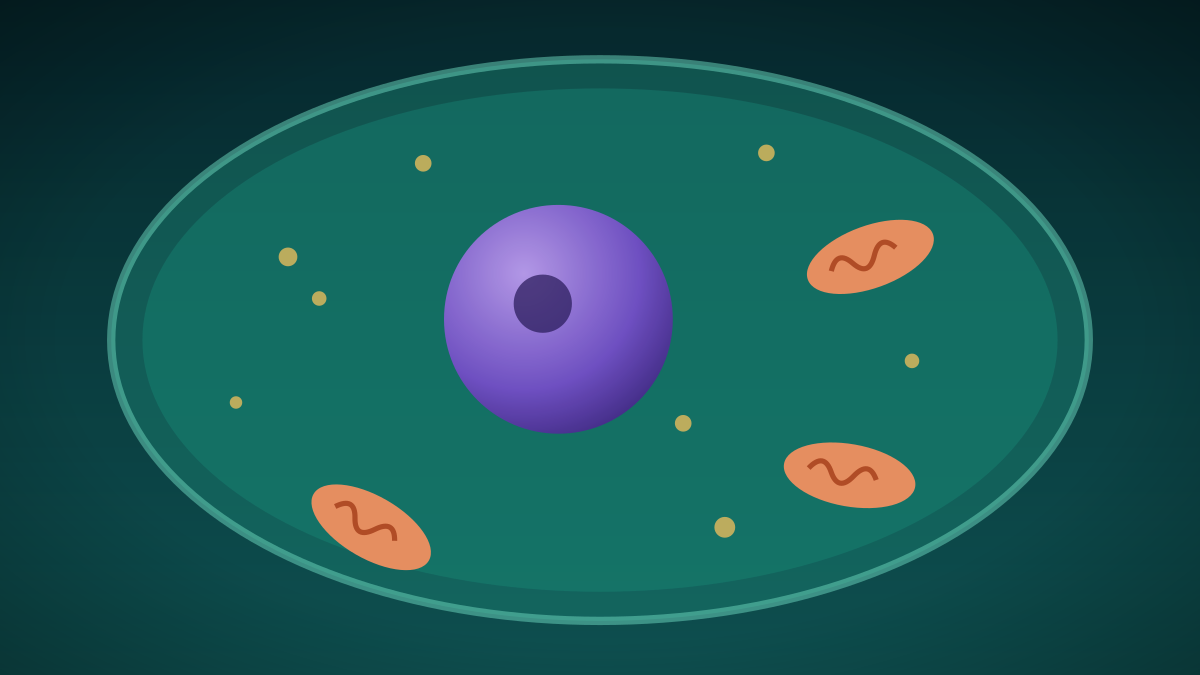
Trillions Strong: Inside the Cell, the Smallest Unit of Life
Every living thing is made of cells, from lone bacteria to the trillions in a human body. A look at the two main cell types and the tiny structures inside them.
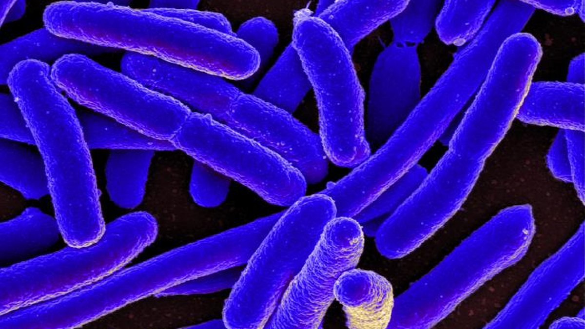
Outnumbered? The Microbes That Live On and In the Human Body
Bacteria, fungi, viruses and other microbes live on skin and in the gut, and researchers have spent years mapping them. Here is what major health agencies say about them.
References
- BC Ministry of Education. Life Sciences 11 (BC curriculum). Accessed October 7, 2026.
- OpenStax. Biology 2e: Studying Cells. Accessed October 7, 2026.
- OpenStax. Biology 2e: Prokaryotic Cells. Accessed October 7, 2026.
- OpenStax. Biology 2e: Eukaryotic Cells. Accessed October 7, 2026.
- OpenStax. Biology 2e: Eukaryotic Origins. Accessed October 7, 2026.
- OpenStax. Biology 2e: Prokaryotic Diversity. Accessed October 7, 2026.
- OpenStax. Biology 2e: Chapter 16 introduction (gene expression). Accessed October 7, 2026.
- NCBI Bookshelf. Molecular Biology of the Cell: Cell differentiation and gene expression. Accessed October 7, 2026.
These lessons follow the content areas listed in the British Columbia curriculum. They are study material written for this site and are not an official document. The official curriculum is the authority on what each course requires. Lessons are general education, not medical advice.