
Metabolism, ATP and Enzymes: How Cells Capture and Use Energy
Catabolism and anabolism, ATP production by glycolysis, the Krebs cycle and the electron transport chain, and how enzymes work and are regulated.
🎯 By the end of this lesson
- Distinguish catabolic from anabolic reactions and give two examples of each.
- Describe how ATP stores and releases energy and name the three kinds of cell work it powers.
- Explain oxidation and reduction and the role of NAD and FAD as electron carriers.
- Outline glycolysis, the Krebs cycle and the electron transport chain, stating location and main products of each.
- Explain why fermentation occurs and what limits its use.
- Describe how an enzyme lowers activation energy and compare the lock-and-key and induced-fit models.
- Predict how temperature, pH and substrate concentration change enzyme activity.
- Explain competitive inhibition, allosteric regulation, feedback inhibition and zymogen activation with examples.
1Overview
A sprinter explodes off the blocks, a student concentrates on a difficult problem, a hair grows a fraction of a millimetre overnight: each involves thousands of chemical reactions running in the right place at the right moment. The sum of all of them is called metabolism. This lesson explains the two directions metabolism can run, the molecule that carries its energy, the stepwise way glucose is dismantled to make that molecule, and the protein catalysts called enzymes that make every step fast enough for life.
2Two directions: catabolism and anabolism
Catabolic reactions break large organic molecules into smaller ones and release energy that was stored in their chemical bonds. Digestion of starch, breakdown of glucose and breakdown of fat for fuel are catabolic. Anabolic reactions do the reverse: they join small molecules into larger ones and require an input of energy. Building protein from amino acids, storing glucose as glycogen and laying down new bone matrix are anabolic.
In terms of Lesson 1, catabolism is mostly hydrolysis and oxidation, and anabolism is mostly dehydration synthesis. The two are linked by a balance sheet. If catabolism releases more energy than anabolism and other work use, the excess is stored, largely as fat. If it releases less, the body draws on stored energy.
Hormones tilt the balance. Catabolic hormones include cortisol (released in stress, raises blood glucose), glucagon (breaks down liver glycogen) and epinephrine. Anabolic hormones include insulin, growth hormone and IGF, and the sex hormones testosterone and estrogen, which support muscle, bone and fat deposition. Lesson 7 returns to these.
Catabolism and anabolism are not rival teams. They are one coordinated network. Catabolism supplies both the ATP and the raw parts that anabolism uses to build and repair.
3ATP: production and use
Cells cannot burn glucose directly in a single step; the energy would be released as useless heat. Instead they capture it in small packets in ATP (adenosine triphosphate). The molecule is a nucleotide: adenine, ribose and three phosphate groups. The bond between the second and third phosphate is the high-energy bond. Hydrolyzing it gives ADP and inorganic phosphate (Pi) plus energy that can be coupled to work; adding the phosphate back (phosphorylation) recharges the molecule.
Only about 40% of the energy released by catabolism is captured in ATP. The remaining roughly 60% is released as heat, which helps explain why working muscle makes the body warm and why shivering raises temperature. ATP is used for three broad categories of work:
- Mechanical work: muscle contraction, movement of chromosomes and cilia.
- Transport work: active transport such as the sodium-potassium pump, which consumes most of a neuron's ATP (Lesson 3).
- Chemical work: driving the anabolic reactions that build macromolecules.
Phosphorylation also regulates proteins
Cells can transfer phosphate from ATP to other molecules. When glucose enters a cell it is phosphorylated, which traps it inside, and phosphorylation of proteins can switch them on or off. In this way ATP is both an energy carrier and a signalling tool.
4Redox reactions and electron carriers
Most energy transfer in metabolism involves moving electrons, usually as hydrogen atoms (an electron plus a proton). Oxidation is the loss of an electron and reduction is the gain of one; the two always occur together, so the electron donor is oxidized while the recipient is reduced. Enzymes catalyze these steps, and helper molecules called coenzymes accept the hydrogen atoms. The two most common are NAD and FAD; their reduced forms, NADH and FADH2, are shuttles that carry energy to the place where most ATP is made.
Energy is not "made" in cells. Energy is converted from one form to another (chemical bond energy in glucose to chemical bond energy in ATP, plus heat). The total is conserved. "Producing ATP" means rebuilding ADP into ATP using energy released from food.
5Cellular respiration: how ATP is made from glucose
Glucose is the main fuel for ATP production. Its complete aerobic breakdown is called cellular respiration and has three stages.
- Glycolysis (cytoplasm). One glucose is first phosphorylated (using 1 ATP, trapping it in the cell) and then, after a second ATP is invested, split into two three-carbon pieces. Each piece is oxidized, reducing NAD+ to NADH, and ATP is made by transferring phosphate to ADP. The products are 2 pyruvate, 4 ATP (a net of 2 after the 2 invested) and 2 NADH. Glycolysis needs no oxygen.
- Pyruvate oxidation and the Krebs (citric acid) cycle (mitochondrial matrix). Pyruvate enters the mitochondrion and is converted to two-carbon acetyl CoA, releasing CO2 and making NADH. Acetyl CoA combines with oxaloacetate to form citrate, and the cycle regenerates oxaloacetate while releasing more CO2. Each turn yields 3 NADH, 1 FADH2 and 1 ATP (through GTP); because one glucose gives two pyruvate, the totals per glucose are doubled.
- Electron transport chain and oxidative phosphorylation (inner mitochondrial membrane). NADH and FADH2 pass electrons along a series of protein complexes. The energy released pumps H+ into the space between the membranes, building a gradient. The H+ then flows back through ATP synthase, which spins like a turbine and attaches phosphate to ADP. Oxygen is the final electron acceptor, combining with electrons and H+ to make water. If oxygen is absent, electron flow stops.
NADH yields about 3 ATP and FADH2 about 2, because FADH2 enters the chain at a later point. The OpenStax textbook gives a net of 36 ATP per glucose for the full aerobic pathway; the large majority comes from the electron transport chain, which is why oxygen supply matters so much.
| Stage | Location | Oxygen needed? | Main products |
|---|---|---|---|
| Glycolysis | cytoplasm | no | 2 pyruvate, net 2 ATP, 2 NADH |
| Krebs cycle | mitochondrial matrix | indirectly (needs NAD+ regenerated) | NADH, FADH2, CO2, a little ATP |
| Electron transport chain | inner mitochondrial membrane | yes | most of the ATP; H2O |
When oxygen is short: fermentation
During intense exercise, muscles use ATP faster than oxygen can be delivered. Glycolysis continues, and pyruvate is converted to lactic acid. This regenerates the NAD+ that glycolysis needs, so ATP production can continue for seconds to a few minutes. Red blood cells have no functional mitochondria and always rely on this route. Lactate diffuses into the blood and travels to the liver, where it can be converted back to pyruvate or glucose (the Cori cycle).
A sprinter runs 200 metres. Stored ATP and creatine phosphate give the first seconds; glycolysis then supplies ATP quickly but inefficiently (net 2 ATP per glucose), and lactic acid accumulates. A distance runner relies instead on aerobic respiration in many mitochondria, which gets far more ATP from each glucose but more slowly. The difference in yield explains why sprints are short and endurance events are paced.
Other fuels and gluconeogenesis
Triglycerides are used mainly for energy through beta-oxidation, and proteins are broken to amino acids that can be rebuilt into proteins or used for ATP. The Krebs cycle supplies starting materials for breaking down proteins and fats. Excess glucose is stored as glycogen in liver and skeletal muscle, or converted to fat. During fasting the liver makes new glucose from pyruvate, lactate, glycerol or amino acids by gluconeogenesis, because the brain and some other organs depend heavily on glucose.
6Fed and fasting: how the body switches between building and burning
Metabolism is not constant; it shifts with the time since the last meal, and hormones direct the switch.
- Fed state (after a meal). Rising blood glucose stimulates beta cells of the pancreas to release insulin. Insulin promotes glucose uptake, glycolysis, storage of glucose as glycogen (glycogenesis) and the building of fats and proteins. The state is mainly anabolic.
- Fasting state (between meals). Falling glucose lowers insulin and raises glucagon. Glucagon stimulates the liver to break glycogen into glucose (glycogenolysis) and to make new glucose (gluconeogenesis), and it stimulates the breakdown of fat. Cortisol and epinephrine push in the same direction during stress. The state is mainly catabolic.
The outcome is that blood glucose stays within a narrow range even though meals are irregular, which matters because neurons and red blood cells depend almost entirely on glucose. The mechanism is the negative feedback loop described in Lesson 3 and examined in detail in Lesson 7.
| Hormone | Source | Metabolic direction | Key effect |
|---|---|---|---|
| Insulin | pancreatic beta cells | anabolic | glucose uptake and glycogen storage |
| Glucagon | pancreatic alpha cells | catabolic | liver glycogen breakdown, raises glucose |
| Cortisol | adrenal cortex | catabolic | gluconeogenesis in prolonged stress |
| Epinephrine | adrenal medulla | catabolic | rapid glucose release in fight or flight |
| Growth hormone, IGF | pituitary, liver | anabolic | growth of bone and tissues |
| Testosterone, estrogen | gonads | anabolic | muscle, bone and fat deposition |
When cortisol is in excess (Cushing syndrome), weight gain concentrates in the trunk and face, blood glucose rises and muscles weaken. When cortisol is deficient (Addison disease), blood pressure falls and fatigue and salt cravings appear, and lifelong replacement is needed. These conditions show how a single catabolic hormone supports normal metabolism only within a narrow range.
Metabolism and body heat
Because about 60% of the energy released in catabolism escapes as heat, metabolism is the body's heater. Thyroid hormones raise the basal metabolic rate, increasing ATP production and oxygen use and releasing more heat (the calorigenic effect). An overactive thyroid therefore causes heat intolerance, sweating and weight loss, whereas an underactive thyroid causes cold intolerance and weight gain. Shivering, in which muscles use ATP to contract without moving the body, is a deliberate way of turning ATP into heat when core temperature falls.
Per glucose, glycolysis produces 4 ATP but invests 2, so the net is 2 ATP. If a textbook states a net of 36 ATP for complete aerobic respiration, then glycolysis supplies a small fraction and the rest comes from the Krebs cycle and the electron transport chain (some of it being spent shuttling NADH into the mitochondria, which is why different books report slightly different totals). In fermentation the net stays at 2 ATP, so the same muscle must burn many times more glucose to produce the same ATP without oxygen.
7Enzymes: biological catalysts
Left alone, most reactions in the body would proceed far too slowly at 37°C. A catalyst speeds a reaction without being consumed, and in the body the important catalysts are enzymes, which are proteins (a few are RNA). Each reaction needs a minimum energy to begin, the activation energy. Enzymes lower it.
An enzyme has an active site, a small pocket whose shape and chemistry fit a particular substrate. Two models describe the fit.
- Lock-and-key model: the substrate fits the rigid active site exactly, like a key in a lock. It explains specificity.
- Induced-fit model: the active site is flexible and adjusts slightly as the substrate binds, like a glove moulding to a hand. This is the model favoured by current textbooks because it also explains how binding strains the substrate bonds and lowers activation energy.
Each enzyme typically catalyzes one reaction or one type of reaction. Many need a non-protein helper: cofactors (often metal ions) or coenzymes (organic molecules such as NAD and FAD, which are derived from vitamins).
8What affects and regulates enzymes
Because an enzyme is a protein whose function depends on shape (Lesson 1), conditions that change shape change activity.
| Factor | Effect |
|---|---|
| Temperature | Rate rises with warmth because particles collide more often, until extreme heat denatures the enzyme and activity collapses. |
| pH | Each enzyme has an optimum. Pepsin works in the acid stomach (pH roughly 1.5 to 3.5), while pancreatic enzymes work in the slightly alkaline small intestine. |
| Substrate concentration | More substrate raises the rate until all active sites are occupied (saturation). |
| Enzyme concentration | More enzyme gives a faster rate when substrate is plentiful. |
Cells also regulate enzymes deliberately, which is how metabolism responds to need.
- Competitive inhibition: a molecule resembling the substrate occupies the active site and blocks it.
- Allosteric regulation: a regulator binds a different site and changes the enzyme's shape, switching it up or down.
- Feedback inhibition: the end product of a pathway inhibits an early enzyme, so the cell stops making more than it needs. This is negative feedback at the molecular scale (Lesson 3).
- Inactive precursors (zymogens): some enzymes are made as inactive forms and activated where needed. Pepsinogen is activated to pepsin by stomach acid, and the pancreas secretes trypsinogen and other protein-digesting enzymes inactive, to prevent digesting the pancreas itself.
- Gene-level control and hormones: insulin and glucagon change which enzymes of glucose metabolism are active, and the cell makes more or fewer copies of an enzyme by changing gene expression (Lesson 4).
In real life. Many drugs are enzyme inhibitors. Aspirin and ibuprofen reduce production of prostaglandins by blocking the enzymes that make them. Enzyme tests are also used in diagnosis. Lactose intolerance, in which the enzyme that hydrolyzes milk sugar is deficient, shows what happens when one enzyme is missing.
Metabolism is therefore a controlled, enzyme-driven network. Its currency is ATP, its main fuel is glucose, its regulation is largely feedback, and it supports everything the later lessons describe, from nerve impulses to muscle contraction to digestion.
🔑Key terms
?Quick check
Try each question first, then reveal the answer.
1. Classify each as anabolic or catabolic: (a) building glycogen from glucose, (b) hydrolysis of starch, (c) forming a peptide bond.
Building glycogen and forming a peptide bond are anabolic because they join small molecules into larger ones using energy. Hydrolysis of starch is catabolic because it breaks a large molecule down and releases energy.
2. Why is ATP called an energy carrier and not an energy store?
Cells hold little ATP at a time; it is continuously hydrolyzed to ADP and phosphate to release energy and continuously rebuilt using energy from catabolism, so it moves energy between reactions rather than keeping it in reserve.
3. Where in the cell does each stage of aerobic respiration occur, and which stage makes most of the ATP?
Glycolysis occurs in the cytoplasm, the Krebs cycle in the mitochondrial matrix and the electron transport chain on the inner mitochondrial membrane. The electron transport chain with ATP synthase makes most of the ATP.
4. What would happen to ATP production in the mitochondria if oxygen were absent, and what would the muscle do instead?
Electrons would have no final acceptor, so the electron transport chain would stop and most ATP production would cease. Glycolysis would continue with pyruvate converted to lactic acid, giving a small amount of ATP for a short time.
5. Explain how an enzyme speeds a reaction without being used up.
The enzyme binds the substrate at its active site and lowers the activation energy needed to form products. After the products are released the enzyme is unchanged and can bind another substrate.
6. How does the induced-fit model differ from the lock-and-key model?
Lock-and-key treats the active site as a rigid shape that matches the substrate exactly. Induced fit says the active site changes shape slightly when the substrate binds, which also strains bonds in the substrate and helps lower activation energy.
7. A person with a fever of 41 degrees Celsius is at risk. Explain using enzyme properties.
Enzymes are proteins that depend on their folded shape. Very high temperature can denature them, changing the active site so they can no longer catalyze reactions, which disrupts metabolism.
8. Describe how feedback inhibition prevents waste in a biosynthetic pathway.
The final product of the pathway binds an early enzyme and reduces its activity. When the product is abundant, production slows; when it is scarce, inhibition lifts and production resumes.
BC curriculum content covered in this lesson
- Metabolism and enzymes: anabolism and catabolism
- Metabolism and enzymes: ATP production and use
- Metabolism and enzymes: enzyme models (lock-and-key, induced fit)
- Metabolism and enzymes: enzyme regulation
Related articles
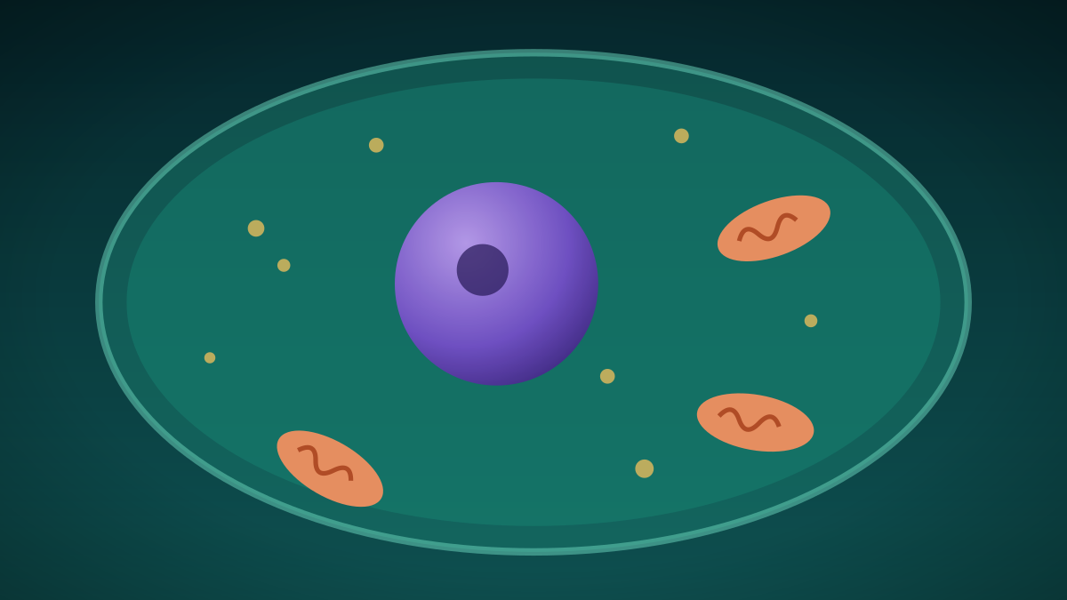
Trillions Strong: Inside the Cell, the Smallest Unit of Life
Every living thing is made of cells, from lone bacteria to the trillions in a human body. A look at the two main cell types and the tiny structures inside them.
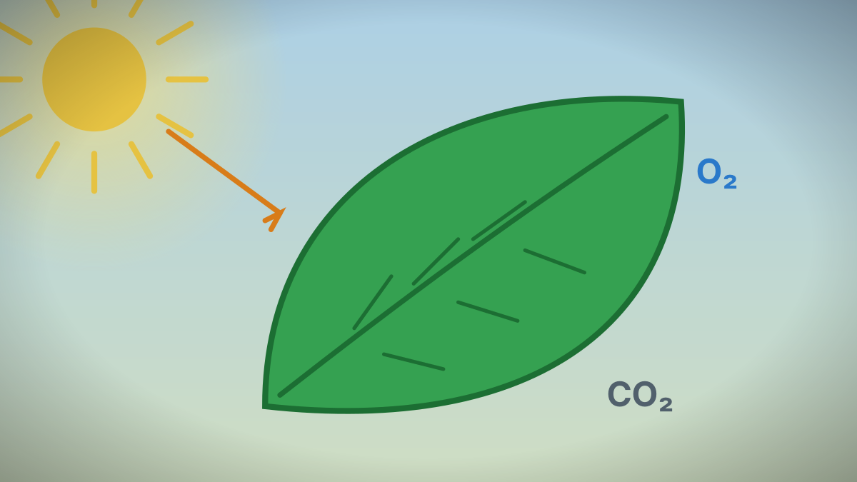
How Photosynthesis Turns Sunlight Into Sugar, Step by Step
Photosynthesis is a two-stage chemical process in chloroplasts that converts light, water and carbon dioxide into sugar and oxygen. Textbooks and ocean scientists explain how it feeds nearly all life.
References
- OpenStax. Anatomy and Physiology 2e, 24.1 Overview of Metabolic Reactions. Accessed October 7, 2026.
- OpenStax. Anatomy and Physiology 2e, 24.2 Carbohydrate Metabolism. Accessed October 7, 2026.
- OpenStax. Anatomy and Physiology 2e, 2.3 Chemical Reactions. Accessed October 7, 2026.
- OpenStax. Anatomy and Physiology 2e, 2.5 Organic Compounds Essential to Human Functioning. Accessed October 7, 2026.
- OpenStax. Anatomy and Physiology 2e, 23.4 The Stomach. Accessed October 7, 2026.
- OpenStax. Anatomy and Physiology 2e, 23.6 Accessory Organs in Digestion. Accessed October 7, 2026.
- BC Ministry of Education and Child Care. Anatomy and Physiology 12 (curriculum). Accessed October 7, 2026.
These lessons follow the content areas listed in the British Columbia curriculum. They are study material written for this site and are not an official document. The official curriculum is the authority on what each course requires. Lessons are general education, not medical advice.