
Mitosis: How One Cell Makes Two Identical Cells
Step-by-step mitosis and cytokinesis, why daughter cells are identical, what mitosis does in a body, and what happens when control is lost (cancer).
🎯 By the end of this lesson
- Students should be able to define mitosis and distinguish it from cytokinesis.
- Students should be able to describe what happens to the chromosomes and nucleus in prophase, prometaphase, metaphase, anaphase and telophase.
- Students should be able to identify the stage of mitosis in a diagram or micrograph.
- Students should be able to compare cytokinesis in animal and plant cells.
- Students should be able to track chromosome and chromatid numbers through mitosis and explain why daughter cells are identical.
- Students should be able to explain the role of the spindle checkpoint.
- Students should be able to explain how failure of cell cycle control can lead to a tumour.
- Students should be able to use cell counts to estimate the relative time spent in each stage.
1One nucleus becomes two
Right now, somewhere in a living body, cells are finishing a copy of their DNA and then splitting it perfectly in two. The result is two cells that are genetically identical to the parent. The process that shares the copied chromosomes is mitosis.
The BC curriculum defines it simply: mitosis is the process through which pre-existing cells make two identical copies of themselves. That definition hides a lot of choreography. Chromosomes must condense, line up, split and travel, all without a single one being lost. This lesson follows that choreography step by step.
Mitosis divides the nucleus so that each new nucleus gets one complete set of chromosomes. Cytokinesis then divides the cytoplasm. Together they turn one cell into two genetically identical cells. DNA copying (S phase) happens before mitosis, during interphase.
Mitosis is easier to follow with a model cell. The diagrams here use a cell with only four chromosomes: two coloured one way and two another, to show that chromosomes come in homologous pairs (one set from each parent). A human cell has 46, but the steps are the same.
2The stages of mitosis, in order
Mitosis is a continuous process, but scientists divide it into named stages so that it is easier to talk about. The stages blend into one another; a real cell does not stop at the boundaries.
Before mitosis: interphase and DNA copying
By the time mitosis begins, the S phase has already copied every chromosome. Each chromosome consists of two sister chromatids joined at the centromere. The centrosome (the organizer of the spindle) has also been duplicated. If mitosis began without this preparation, the daughter cells would receive only half of the instructions.
The five phases
- Prophase. The loosely spread chromatin winds up tightly into visible chromosomes. The nucleolus disappears, and the nuclear envelope breaks into small vesicles. The two centrosomes move to opposite ends (poles) of the cell, and the spindle, a framework of tiny tubes called microtubules, grows between them.
- Prometaphase. The remnants of the nuclear envelope fragment further. Each sister chromatid develops a protein structure called a kinetochore at its centromere. Spindle fibres from opposite poles attach to the kinetochores, so that each of the two sister chromatids faces a different pole. (Many introductory courses fold this stage into prophase or metaphase.)
- Metaphase. The spindle fibres tug the chromosomes until they line up along the cell's middle, an imaginary plane called the metaphase plate. Chromosomes are at their most condensed here, which is why metaphase is the stage most often used for pictures of chromosomes. A checkpoint confirms that every chromosome is properly attached before the cell continues.
- Anaphase. The proteins holding sister chromatids together are broken down, and the sister chromatids separate at the centromere. Each chromatid is now counted as a full chromosome and is pulled towards the pole its fibre leads to. The cell also elongates as the poles move apart.
- Telophase. The chromosomes reach the poles and unwind back into loose chromatin. The spindle breaks down, and a nuclear envelope re-forms around each set of chromosomes. The cell now has two nuclei.
| Stage | What happens to the chromosomes | What happens to the nucleus and spindle |
|---|---|---|
| Prophase | Condense and become visible; each is two sister chromatids | Envelope breaks up; spindle begins to form |
| Prometaphase | Attach to spindle fibres at kinetochores | Envelope fragments completely |
| Metaphase | Line up in the middle of the cell | Spindle fully formed |
| Anaphase | Sister chromatids separate and move to opposite poles | Cell lengthens |
| Telophase | Reach the poles and unwind | Two nuclear envelopes re-form; spindle disappears |
“Interphase is when the cell is resting.” Interphase is the opposite of resting. The cell grows, makes the materials it needs and copies its DNA. Only certain cells that have left the cycle (G0) can be described as resting. Mitosis itself is the short, dramatic part.
3Cytokinesis: dividing the rest of the cell
Mitosis moves the chromosomes, but a cell is more than a nucleus. The cytoplasm, organelles and membrane must also be divided. This is cytokinesis. It usually starts in late anaphase and finishes after telophase.
In animal cells
A ring of protein fibres (actin) forms just inside the cell membrane around the middle of the cell, where the metaphase plate was. As the ring tightens, it pulls the membrane inward, creating a groove called the cleavage furrow. The furrow deepens until the cell pinches into two. A useful comparison is the drawstring of a bag pulled tight.
In plant cells
Plant cells are surrounded by a stiff wall, so they cannot simply pinch. Instead, small membrane-bound packages called vesicles, carrying building materials, gather in the centre of the cell and fuse into a flat structure called the cell plate. The plate grows outward until it reaches the existing walls. The materials are then used to build a new wall between the two daughter cells, and the vesicle membranes become the new cell membranes.
| Feature | Animal cell | Plant cell |
|---|---|---|
| Starting structure | Ring of fibres at the middle | Vesicles gathering at the middle |
| How the cell splits | Membrane pinches inward (cleavage furrow) | Cell plate grows outward to the walls |
| Direction of growth | From the outside inward | From the centre outward |
| Cell wall | None to build | New wall forms between daughter cells |
Cytokinesis does not always happen. If it is skipped after mitosis, the result is a single cell with more than one nucleus. Both outcomes show that mitosis (nuclear division) and cytokinesis (cell division) are two separate steps.
4Why the daughter cells are identical
The result of mitosis is two cells with the same number and type of chromosomes as the parent cell. Two features of the process guarantee this.
- The DNA is copied exactly once. The sister chromatids in each chromosome are identical copies.
- The sister chromatids are pulled to opposite poles. Each new cell receives exactly one copy of every chromosome. The spindle checkpoint helps to make sure that no chromosome is attached to only one pole.
A cell in a certain species has 8 chromosomes. It is in G1. How many chromosomes and chromatids does it have at each stage, and how many does each daughter cell have?
- G1: 8 chromosomes, each a single chromatid, so 8 chromatids.
- After S phase and through metaphase: still 8 chromosomes, but each has two sister chromatids, so 16 chromatids.
- Anaphase: sister chromatids separate, so there are briefly 16 separate chromosomes, 8 moving to each pole.
- After cytokinesis: each daughter cell has 8 chromosomes.
The number goes 8, 8, 16, 8. It returns to the starting number because the doubling at anaphase is shared between two cells.
“Identical” refers to the DNA that is passed on. Small copying errors (mutations) can happen, but they are rare, and cells have repair and checkpoint systems to catch many of them. The daughter cells can also differ in other ways, for example in how the cytoplasm is divided.
5Mitosis at work in a body
Mitosis is the main type of cell division used by eukaryotic organisms for growth, repair and replacement. Its jobs in the body include the following.
- Growth. After fertilization, a single cell divides again and again by mitosis. The early divisions produce a ball of smaller and smaller cells, and later divisions build tissues and organs.
- Repair. When skin is scraped, cells near the wound divide to replace those that were lost.
- Replacement. Many cells wear out and are replaced by new ones made through mitosis.
- Asexual reproduction. In some single-celled eukaryotes and in many multicellular organisms, mitosis creates new individuals without a partner. This is the topic of the next lesson.
Not every cell keeps dividing. Mature heart muscle cells and nerve cells enter G0 and generally stay there, which is one reason damage to these tissues is hard to repair. Cells in G0 are not dead; they have simply stopped cycling. Other tissues, by contrast, keep making new cells to replace those that wear out.
6When the controls fail: cancer
Because mitosis produces more cells, it must be tightly controlled. Cells normally divide only when needed, and checkpoints pause the cycle when something is wrong. Cancer is a group of diseases caused by uncontrolled cell growth.
The story usually begins with DNA copying errors. Even with checkpoints in place, a small share of errors in S phase may get through and be passed on to daughter cells. Errors can also be caused by damage to DNA. If the errors build up in genes that control the cycle, the controls weaken. Two kinds of genes matter most.
- Genes for “accelerators”. Normal genes called proto-oncogenes code for proteins that push the cell forward. When altered so that they are overactive, they are called oncogenes and push the cell through the cycle too fast.
- Genes for “brakes”. Tumour suppressor genes code for proteins (such as p53) that stop division when something is wrong. When they are damaged, the brakes fail. A damaged form of p53 is found in more than half of human tumour cells.
Cells that no longer respond to the controls can outgrow their neighbours and form a lump called a tumour. In short: the cell cycle and its checkpoints are what stand between a healthy body and runaway growth.
“Cancer is a foreign invader.” Cancer cells come from the body's own cells that have accumulated changes in the genes controlling the cell cycle. They are the body's own cells dividing without the normal controls.
Reading mitosis under a microscope
Identifying stages in a photograph or slide of dividing cells is a standard skill. A few clues make it manageable.
- Is a nuclear envelope visible? If a clear round nucleus is visible and no distinct chromosomes can be seen, the cell is in interphase.
- Are chromosomes visible as separate threads or rods? If so, and no line-up is visible, the cell is in prophase (or prometaphase).
- Are chromosomes in a line across the middle? Metaphase.
- Are two groups of chromosomes moving apart? Anaphase, often with V-shapes pointing toward the poles.
- Are there two separate groups at opposite ends, with new nuclear envelopes or a pinching middle? Telophase and cytokinesis.
In a field of view of 50 cells from a rapidly growing tissue, a student finds 39 in interphase, 6 in prophase, 2 in metaphase, 1 in anaphase and 2 in telophase. Interphase accounts for 39/50 = 78% of the cells. If the whole cycle is assumed to take 20 hours, interphase lasts about 15.6 hours. Prophase is the longest of the mitotic stages (6/50 = 12%, or about 2.4 hours), while anaphase is the shortest (1/50 = 2%, or about 0.4 hours). The numbers are invented for practice, but the method is real: more cells in a stage means the stage lasts longer.
This links back to the idea from the previous lesson that frozen snapshots of many cells can be used to estimate how long each stage takes.
What if something goes wrong during division?
The spindle checkpoint exists because the stakes are high. Consider what would happen if a single sister-chromatid pair were attached to spindle fibres from only one pole. At anaphase, both chromatids could be dragged to the same side. One daughter cell would then have an extra chromosome and the other would be missing one. Missing or extra chromosomes mean missing or extra copies of many genes, which usually upsets the way a cell works.
This is why the checkpoint holds the cell in metaphase until every chromosome is attached to fibres from opposite poles. The cell treats an unattached chromosome as a reason to wait. Only when everything is in place does it let anaphase start. Thinking this way, the checkpoint is a quality-control step, similar to a pilot running through a checklist before takeoff.
The accuracy of mitosis comes from three things working together: exact DNA copying in S phase, a spindle that pulls sister chromatids to opposite poles, and checkpoints that stop the process whenever something is not right.
Modelling mitosis
A physical or drawn model is a good way to test whether the stages make sense. A simple model can be built with two colours of string or pipe cleaner, using two pairs of chromosomes as in the diagram.
- Lay out four single strands: two of one colour (a homologous pair) and two of another colour (a second pair).
- Show S phase by adding a matching copy next to each strand and twisting each pair together at the middle (the centromere). There are still four chromosomes, but each has two chromatids.
- Line the four joined pairs along a line down the middle of the table (metaphase).
- Pull each joined pair apart so one strand goes to each side (anaphase). Count the strands on each side.
- Draw a circle round each group of strands (telophase and cytokinesis).
The model has limits: real chromosomes are not strings, and real cells do not move chromosomes by hand. Evaluating what a model does and does not show is itself part of scientific thinking. Even so, each side ends up with four strands that match the original set, which captures the main outcome of mitosis.
7Summary
- Mitosis follows interphase and divides the nucleus into two identical nuclei: prophase, (prometaphase), metaphase, anaphase, telophase.
- Cytokinesis divides the cytoplasm. Animal cells pinch (cleavage furrow); plant cells build a cell plate.
- Because DNA is copied once and sister chromatids are shared equally, the two daughter cells are genetically identical to the parent and to each other.
- Mitosis supports growth, repair and replacement, and, in many organisms, reproduction.
- Failure of cell cycle controls can lead to uncontrolled division and tumours (cancer).
Mitosis makes identical copies. The next lesson shows how organisms use it to reproduce on their own. After that, a different kind of division, meiosis, is needed to produce sex cells.
🔑Key terms
?Quick check
Try each question first, then reveal the answer.
1. State the difference between mitosis and cytokinesis.
Mitosis divides the nucleus into two identical sets of chromosomes. Cytokinesis divides the cytoplasm, producing two separate cells.
2. Put these events in order: sister chromatids separate, nuclear envelopes re-form, chromosomes line up in the middle, chromosomes condense.
Chromosomes condense (prophase), line up in the middle (metaphase), sister chromatids separate (anaphase), nuclear envelopes re-form (telophase).
3. A micrograph shows a cell with two groups of V-shaped chromosomes at opposite ends and an elongated cell. Name the stage and justify the answer.
Anaphase. Sister chromatids have separated and are being pulled to opposite poles, and the cell has lengthened as the poles move apart.
4. Compare how animal cells and plant cells complete cytokinesis.
An animal cell is pinched in two by a contracting ring that forms a cleavage furrow. A plant cell builds a cell plate across its middle that grows outward and becomes a new cell wall.
5. A cell with 10 chromosomes enters mitosis. How many chromatids are present at metaphase, and how many chromosomes does each daughter cell have?
There are 20 chromatids at metaphase because each of the 10 chromosomes has two sister chromatids. Each daughter cell has 10 chromosomes.
6. Why are the two daughter cells genetically identical to the parent cell?
DNA was copied exactly once, and the sister chromatids were pulled to opposite poles, so each daughter cell received one copy of every chromosome.
7. Predict what would happen if a pair of sister chromatids were both pulled to the same pole in anaphase, and name the checkpoint that normally prevents this.
One daughter cell would have an extra chromosome and the other would be missing one. The spindle checkpoint in metaphase prevents this by waiting until every chromosome is attached to fibres from opposite poles.
8. Explain how a mutation in a tumour suppressor gene can lead to a tumour.
A tumour suppressor gene codes for proteins that stop division when there is a problem. If the gene is damaged, the brakes fail, so damaged cells keep dividing and can build up into a tumour.
BC curriculum content covered in this lesson
- Mitosis: the process through which pre-existing cells make two identical copies of themselves
- Stages of mitosis (prophase, metaphase, anaphase, telophase) and cytokinesis in animal and plant cells
- Mitosis for growth, repair and replacement; cell cycle control and cancer
Related articles
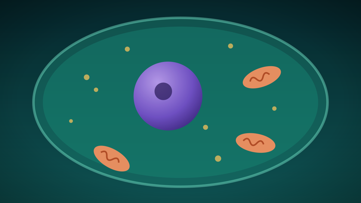
Trillions Strong: Inside the Cell, the Smallest Unit of Life
Every living thing is made of cells, from lone bacteria to the trillions in a human body. A look at the two main cell types and the tiny structures inside them.
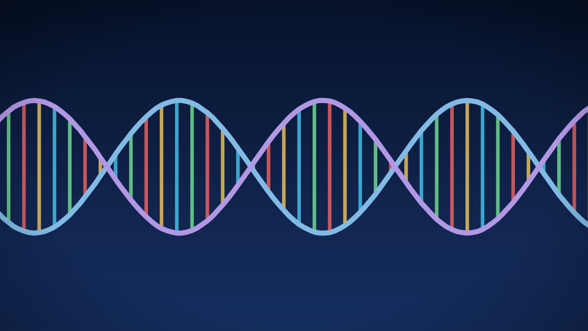
What Is DNA? The Molecule That Carries Your Instructions
DNA is the molecule that carries genetic information. A short guide to its structure and how its letters pair.
References
- BC Ministry of Education and Child Care. Science 9 curriculum (Big Ideas, Content, Curricular Competencies). Accessed October 7, 2026.
- OpenStax. Biology 2e, 10.1 Cell Division. Accessed October 7, 2026.
- OpenStax. Biology 2e, 10.2 The Cell Cycle. Accessed October 7, 2026.
- OpenStax. Biology 2e, 10.3 Control of the Cell Cycle. Accessed October 7, 2026.
- OpenStax. Biology 2e, 10.4 Cancer and the Cell Cycle. Accessed October 7, 2026.
These lessons follow the content areas listed in the British Columbia curriculum. They are study material written for this site and are not an official document. The official curriculum is the authority on what each course requires. Lessons are general education, not medical advice.