
The Nervous System: Neurons, Signals, Brain and Autonomic Control
Neuron structure, action potentials and synapses, the organization of the brain and spinal cord, autonomic control of organs and the nervous system's role in homeostasis.
🎯 By the end of this lesson
- Distinguish the CNS from the PNS and the somatic, autonomic and enteric divisions.
- Label the parts of a neuron and explain the role of myelin in conduction.
- Describe the sequence of events in an action potential and relate each to ion movement.
- Explain the all-or-none principle, the refractory period and saltatory conduction.
- Outline the steps of transmission at a chemical synapse and distinguish EPSPs from IPSPs.
- Match major brain regions to their functions and trace a simple reflex arc.
- Compare the sympathetic and parasympathetic divisions in anatomy, transmitters and effects.
- Explain how the nervous system acts as sensor and effector in at least three homeostatic loops.
1Overview
Touching a hot pan produces a withdrawn hand before the word "ouch" is even formed. In less than a second, receptors in the skin fire, signals race along nerves to the spinal cord, a motor command returns to muscle and the hand moves. That sequence is the nervous system at its simplest: detect, integrate, respond. The nervous system also runs the heartbeat while a person sleeps, adjusts the pupils to a change in light and stores memories. This lesson covers its cells, its electrical and chemical signals, its organization and its role in homeostasis.
2Organization of the nervous system
The nervous system has three basic functions: sensation (detecting stimuli from outside and inside the body), integration (combining sensory input with memory, emotion and context to choose a response) and response (motor output to skeletal, cardiac and smooth muscle, and to glands).
| Division | Parts | Role |
|---|---|---|
| Central nervous system (CNS) | brain and spinal cord | integration and command |
| Peripheral nervous system (PNS) | everything else: nerves and ganglia | carries information to and from the CNS |
| Somatic nervous system | part of the PNS | conscious perception and voluntary control of skeletal muscle; also reflexes |
| Autonomic nervous system (ANS) | part of the PNS; sympathetic and parasympathetic | involuntary control of smooth muscle, cardiac muscle and glands to maintain homeostasis |
| Enteric nervous system | about 100 million neurons in the wall of the digestive tract | controls digestive muscle and glands; can work without the CNS |
3Neurons and glial cells
Nervous tissue contains two cell types. Neurons carry out communication. Glial cells support neurons; for example, astrocytes help buffer the potassium that neurons release. A neuron has a soma (cell body), branched dendrites that receive most input, and a single axon that carries the signal to its target.
Many axons are wrapped in myelin, a lipid-rich insulating layer. Myelin gives white matter its colour and speeds conduction. In the CNS, gray matter is regions rich in cell bodies and dendrites and white matter is regions dominated by axons. Names differ by location: a cluster of cell bodies is a nucleus in the CNS but a ganglion in the PNS, and a bundle of axons is a tract in the CNS but a nerve in the PNS.
4The action potential: an electrical signal
A neuron at rest is electrically polarized. The inside of the membrane is about -70 mV relative to the outside. Na+ is about ten times more concentrated outside the cell, and K+ is more concentrated inside. Leakage channels let a little Na+ in and K+ out, and the sodium-potassium pump (Lesson 3) restores the gradients using ATP.
- Threshold. A stimulus depolarizes the membrane. If it reaches about -55 mV, voltage-gated Na+ channels open. A smaller change fails to trigger anything.
- Depolarization. Na+ rushes in, driven by concentration and electrical gradients, making the inside positive, to about +30 mV.
- Repolarization. The Na+ channels inactivate and voltage-gated K+ channels open. K+ leaves and the membrane returns toward rest.
- Hyperpolarization. The K+ channels close slightly late, so the membrane briefly dips below -70 mV before the pump and leakage channels restore the resting state.
The whole event lasts about 2 ms and changes the voltage by about 100 mV (0.1 V). Three properties follow.
- All-or-none: either threshold is reached and a full action potential occurs, or nothing happens. Every action potential peaks at about +30 mV. A stronger stimulus produces more action potentials per second, not larger ones.
- Refractory period: while Na+ channels are inactivated no new action potential can start (absolute); soon after, a stronger-than-usual stimulus is needed (relative). This keeps signals moving in one direction.
- Propagation: in an unmyelinated axon the signal moves continuously along the membrane, which is slower. In a myelinated axon the signal jumps between nodes of Ranvier (saltatory conduction), which is faster. A wider axon also conducts faster.
An action potential is not a current of electrons flowing through the axon like electricity in a wire. It is a wave of changing ion permeability: ions cross the membrane at one patch, which triggers the next patch, and so on. The cell's ATP (through the pump) restores the gradients afterward.
A stimulus raises the membrane from -70 mV to -60 mV. This is a graded potential that does not reach threshold (-55 mV), so no action potential occurs. A second stimulus that raises it from -70 mV to -50 mV does cross threshold, and the neuron fires a full +30 mV spike. A third, much stronger stimulus fires the neuron repeatedly but each spike is the same size. The neuron encodes intensity by frequency.
5Synapses: from electrical to chemical signal
A synapse is the junction where one neuron communicates with another cell. Some synapses are electrical (cells directly connected so ions pass), but the typical one is chemical.
- An action potential reaches the axon terminal and opens voltage-gated Ca2+ channels.
- Ca2+ entering binds proteins on synaptic vesicles, which fuse with the presynaptic membrane and release neurotransmitter by exocytosis.
- The neurotransmitter diffuses across the synaptic cleft and binds receptors on the postsynaptic membrane, like a key in a lock.
- The effect ends when the neurotransmitter is broken down by an enzyme (acetylcholine by acetylcholinesterase), or taken back up by the neuron or by glial cells.
Whether the postsynaptic cell is excited depends on the receptor. An excitatory postsynaptic potential (EPSP) depolarizes the membrane toward threshold; an inhibitory postsynaptic potential (IPSP) hyperpolarizes it away from threshold. A neuron adds its inputs together at the initial segment of the axon. Spatial summation combines inputs from different places, and temporal summation combines repeated inputs from one place that arrive close together. If the total reaches threshold, an action potential fires.
| Neurotransmitter | Where and effect |
|---|---|
| Acetylcholine | neuromuscular junction, autonomic nervous system and brain; excitatory at nicotinic receptors |
| Glutamate | main excitatory transmitter of the brain |
| GABA and glycine | inhibitory, hyperpolarize the postsynaptic cell |
| Dopamine, norepinephrine, serotonin | biogenic amines; effects depend on the receptor type |
| Endorphins, substance P | neuropeptides, often released with another transmitter |
Receptors are either ionotropic (the receptor is itself an ion channel, so the effect is fast) or metabotropic (acting through a G protein and second messenger, so the effect is slower but can alter metabolism or gene expression, and may underlie learning and memory).
In real life. Nicotine binds and activates nicotinic acetylcholine receptors, mimicking acetylcholine. Muscarine from certain mushrooms instead binds muscarinic receptors. Drugs and toxins often act at synapses because a synapse is a place where a molecule can substitute for a transmitter, block a receptor or prevent removal of the transmitter.
6The central nervous system and sensation
From stimulus to perception
Sensation begins at sensory receptors, specialized endings or cells that convert a stimulus into electrical signals. Different receptor types respond to different forms of energy: mechanoreceptors to touch, pressure and stretch, thermoreceptors to temperature, nociceptors to damaging stimuli (pain), chemoreceptors to chemicals such as CO2 and H+, and photoreceptors to light. Receptors inside the body, such as stretch receptors in the walls of the bladder and arteries, supply the information that feedback loops need.
- A stimulus changes the membrane potential of a receptor (a graded potential).
- If the change reaches threshold, an action potential travels along a sensory neuron toward the CNS.
- Synapses pass the signal through the spinal cord or brain stem, usually via the thalamus for conscious senses.
- The cortex interprets the signal; location of the activity (for example the postcentral gyrus for touch or the occipital lobe for vision) determines what is perceived, and the pattern and frequency of action potentials indicate how strong it is.
- If a response is needed, motor neurons carry commands to effectors.
A student grips a very cold glass. Thermoreceptors in the fingers fire; sensory neurons carry the signal to the spinal cord and up to the thalamus and the parietal cortex, so the cold is perceived and located in the hand. At the same time hypothalamic circuits note falling skin temperature and, if the body as a whole is cooling, sympathetic neurons narrow the skin's blood vessels. Perception and homeostasis use the same sensory information in different pathways.
Protecting the CNS
The brain and spinal cord are soft and demand a constant, stable chemical environment. They are protected by the skull and vertebral column and by membranes (meninges) and fluid around them. A specialized barrier also controls what enters brain tissue: capillaries in the brain have tight junctions with no gaps between endothelial cells and a thick basement membrane, forming the blood-brain barrier. Small lipid-soluble molecules can cross, while many substances in the blood are kept out and others, such as glucose, enter by specific carrier proteins (Lesson 3). The barrier is one reason that drug design for the brain is difficult.
| Protective feature | What it does |
|---|---|
| Skull and vertebrae | hard protection against impact |
| Meninges and cerebrospinal fluid | cushion the CNS and provide a stable fluid environment |
| Blood-brain barrier | restricts which substances reach neurons |
| Glial cells | support neurons and buffer ions such as K+ |
The brain
| Region | Main functions |
|---|---|
| Cerebrum: frontal lobe | voluntary movement (precentral gyrus), planning, speech production (Broca's area, usually left) |
| parietal lobe | touch, pain, body position and movement sense |
| occipital lobe | primary vision |
| temporal lobe | hearing and memory |
| Basal nuclei, hippocampus, amygdala | coordination of movement; long-term memory and emotion |
| Thalamus | relays sensory information (except smell) to the cortex |
| Hypothalamus | regulates homeostasis, controls the autonomic nervous system and governs the pituitary |
| Brain stem: midbrain, pons, medulla | medulla controls cardiovascular and respiratory function; the reticular formation supports sleep and wakefulness |
| Cerebellum | compares motor commands with sensory feedback to coordinate movement |
The two cerebral hemispheres are connected by the corpus callosum. The cerebellum accounts for about 10% of brain mass.
The spinal cord and reflexes
The spinal cord has posterior (dorsal) regions that receive sensory input and anterior (ventral) regions that send motor output to skeletal muscle; lateral horns in the thoracic, upper lumbar and sacral regions contain autonomic neurons. White columns carry ascending sensory and descending motor tracts. A reflex is a rapid, automatic response handled largely in the cord, which is why the hand withdraws from a hot pan before the brain registers pain. A reflex arc has a receptor, a sensory neuron, an integration centre in the CNS, a motor neuron and an effector.
A widely repeated claim says people use only 10% of their brain. Imaging that tracks blood flow shows that most of the brain is active during well-defined tasks, and different regions are active at different times. A related site article examines this idea.
7The autonomic nervous system
The autonomic system has two divisions that usually act in opposition.
| Feature | Sympathetic | Parasympathetic |
|---|---|---|
| Nickname | fight or flight | rest and digest |
| Origin | thoracic and upper lumbar spinal cord | brainstem (including the vagus nerve) and sacral cord |
| Ganglia | chain beside the spine and collateral ganglia | near or within the target organ |
| Pre- / postganglionic fibres | short / long | long / short |
| Main postganglionic transmitter | norepinephrine (acetylcholine to sweat glands) | acetylcholine |
| Scope | widespread; one preganglionic neuron can reach 10 to 20 targets | more localized |
| Typical effects | faster heart rate and breathing, blood to skeletal muscle, less blood to digestion, sweating | slower heart (vagus), more digestive activity, pupil, saliva and tear control |
The adrenal medulla acts like a modified sympathetic ganglion: stimulated by preganglionic fibres, it releases epinephrine and norepinephrine into the blood, so the same chemicals act as hormones. Most organs have dual innervation, so their activity reflects the balance of the two divisions. The sympathetic division raises heart rate; the parasympathetic division lowers it. The term "fight or flight" dates from 1915, but the sympathetic system also responds to emotional states, so "fright or freeze" can be included.
8Homeostasis and interdependence
The nervous system is the fast sensor and effector arm of most feedback loops.
- Blood pressure. Baroreceptors in the aorta and carotid arteries report pressure to the medulla, which adjusts heart rate and vessel diameter through the autonomic system (Lesson 8).
- Breathing. The medulla sets the rhythm of breathing and responds to blood CO2 and pH (Lesson 10).
- Temperature. The hypothalamus compares body temperature with its set point and drives sweating, vasodilation, vasoconstriction and shivering (Lesson 3).
- Digestion. Parasympathetic activity increases motility and secretion; sympathetic activity decreases them, and the enteric network coordinates local reflexes (Lesson 11).
- Endocrine control. The hypothalamus links the nervous and endocrine systems through the pituitary, so many neural signals become hormonal responses (Lesson 7).
The nervous system works in milliseconds and the endocrine system in seconds to hours, but neither works alone. The hypothalamus is the meeting point: it is nervous tissue that releases hormones and commands the pituitary gland.
In real life. After a stroke or loss of blood flow to the brain, the extracellular K+ concentration rises, astrocytes lose their buffering ability and the sodium-potassium pump can fail, so neurons cannot maintain their gradients. In Parkinson disease, loss of dopamine-producing neurons in the substantia nigra disturbs the basal nuclei's control of movement; L-DOPA helps because it can cross the blood-brain barrier and be converted into dopamine. Both conditions show that structure, ions, transmitters and blood supply are linked.
🔑Key terms
?Quick check
Try each question first, then reveal the answer.
1. Describe the movement of ions during depolarization and repolarization.
During depolarization voltage-gated Na+ channels open and Na+ rushes into the neuron, making the inside positive. During repolarization the Na+ channels inactivate and K+ channels open, so K+ leaves and the inside becomes negative again.
2. Why does a stronger stimulus not produce a bigger action potential?
Action potentials are all-or-none: once threshold is reached each one reaches the same peak. A stronger stimulus makes the neuron fire more action potentials per second, so intensity is coded by frequency.
3. Why do myelinated axons conduct faster than unmyelinated ones?
Myelin insulates the axon so the action potential regenerates only at the nodes of Ranvier and jumps from node to node (saltatory conduction), rather than at every point along the membrane.
4. List the sequence of events at a chemical synapse from the arrival of an action potential to the end of the signal.
Ca2+ channels open and Ca2+ enters the terminal; vesicles fuse and release neurotransmitter by exocytosis; it binds receptors on the postsynaptic cell; the signal ends when the transmitter is degraded by an enzyme or taken back up.
5. A neuron receives many inputs. Explain how it decides whether to fire.
It adds EPSPs and IPSPs through spatial and temporal summation at the initial segment of the axon. If the net depolarization reaches threshold (about -55 mV) an action potential fires; otherwise it does not.
6. Compare the ganglia and fibre lengths of the sympathetic and parasympathetic divisions.
Sympathetic ganglia lie near the spinal cord, so preganglionic fibres are short and postganglionic fibres are long. Parasympathetic ganglia lie near or within the organ, so preganglionic fibres are long and postganglionic fibres are short.
7. Explain why a person's heart rate rises during a sudden fright and then slows after the danger passes.
Sympathetic activation releases norepinephrine (and epinephrine from the adrenal medulla), raising heart rate. When the threat ends, sympathetic activity falls and parasympathetic (vagal) activity, using acetylcholine, slows the heart again; dual innervation balances the two.
8. Which part of the brain acts as the link between the nervous and endocrine systems, and how?
The hypothalamus. It regulates the autonomic nervous system and homeostasis, and it controls the pituitary gland through releasing hormones and by sending neurosecretory axons to the posterior pituitary.
BC curriculum content covered in this lesson
- Organ systems: nervous system (structure and function)
- Structural and functional interdependence (nervous system with the endocrine, cardiovascular, respiratory and digestive systems)
- Maintenance of homeostasis (nervous system)
Related articles
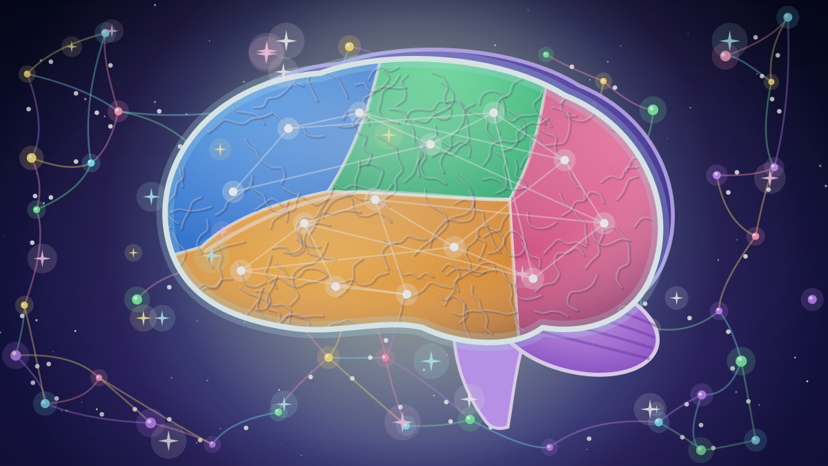
Do People Really Use Only 10 Percent of the Brain?
The claim that only 10 percent of the brain is used is a myth. Imaging, brain-damage evidence and energy costs show activity throughout the brain, and the figure has murky origins.
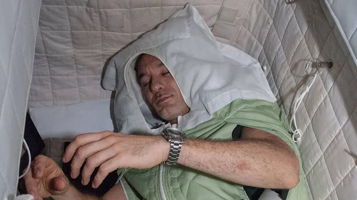
Why Sleep Is Not Downtime: What the Brain and Body Do After Dark
Sleep is an active state in which the brain, heart, hormones and immune system all shift gears. US health agencies also publish recommended hours by age, and their figures differ slightly.
Why Do People Yawn?
Yawning has no settled explanation. Brain cooling, arousal and social signalling are the leading ideas, while the oxygen theory has been dropped. Evidence and limits are summarised.
References
- OpenStax. Anatomy and Physiology 2e, 12.1 Basic Structure and Function of the Nervous System. Accessed October 7, 2026.
- OpenStax. Anatomy and Physiology 2e, 12.4 The Action Potential. Accessed October 7, 2026.
- OpenStax. Anatomy and Physiology 2e, 12.5 Communication Between Neurons. Accessed October 7, 2026.
- OpenStax. Anatomy and Physiology (1e), 13.2 The Central Nervous System. Accessed October 7, 2026.
- OpenStax. Anatomy and Physiology 2e, 15.1 Divisions of the Autonomic Nervous System. Accessed October 7, 2026.
- OpenStax. Anatomy and Physiology 2e, 17.1 An Overview of the Endocrine System. Accessed October 7, 2026.
- OpenStax. Anatomy and Physiology 2e, 23.1 Overview of the Digestive System. Accessed October 7, 2026.
- OpenStax. Anatomy and Physiology 2e, 25.9 Regulation of Fluid Volume and Composition. Accessed October 7, 2026.
- OpenStax. Anatomy and Physiology 2e, 22.3 The Process of Breathing. Accessed October 7, 2026.
- BC Ministry of Education and Child Care. Anatomy and Physiology 12 (curriculum). Accessed October 7, 2026.
These lessons follow the content areas listed in the British Columbia curriculum. They are study material written for this site and are not an official document. The official curriculum is the authority on what each course requires. Lessons are general education, not medical advice.