
The Lymphatic and Immune Systems: Fluid Return and Defence
Structure of the lymphatic system, innate defences and inflammation, adaptive immunity through T and B cells, vaccines, and immune disorders.
🎯 By the end of this lesson
- Describe the pathway of lymph and the roles of nodes, spleen, thymus and tonsils.
- Explain how the lymphatic system supports fluid balance and fat absorption.
- List the barriers and cells of innate immunity and explain their functions.
- Describe the four steps of inflammation.
- Distinguish cell-mediated from humoral immunity and name the major T cell types.
- Explain clonal selection, antibody classes and why a secondary response is faster.
- Compare active and passive immunity and explain how vaccines work.
- Explain how allergy, autoimmune disease and HIV infection represent failures of immune balance.
1Overview
The body is under constant attack. Bacteria on the skin, viruses in the air and abnormal cells arising from within all threaten the internal environment, yet most are neutralized without the person ever noticing. Two linked systems manage this. The lymphatic system drains and filters the fluid that leaks from capillaries and houses the cells of defence. The immune system, built from these cells and their chemicals, recognizes and destroys threats and remembers them. A related article describes the immune system in general; this lesson adds the structure, mechanisms and links to homeostasis.
2Structure of the lymphatic system
The lymphatic system has three roles: returning tissue fluid to the blood, absorbing dietary fat from the intestine, and providing a base for immune cells.
- Lymph and capillaries. Interstitial fluid that enters the lymphatic system is called lymph. Lymphatic capillaries are blind-ended tubes made of overlapping endothelial cells that act as one-way flaps: fluid enters, but cannot flow back. They are absent from the central nervous system, bone marrow, bones, teeth and the cornea.
- Vessels and ducts. Larger vessels resemble veins and have valves. Lymph is not pumped by the heart; it moves through skeletal muscle contraction, body movement and breathing, with valves keeping it flowing toward the heart. The right lymphatic duct drains the upper right body into the right subclavian vein, and the thoracic duct drains the rest into the left subclavian vein.
- Lacteals. In the small intestine lymph capillaries called lacteals absorb dietary fats and fat-soluble vitamins as a milky fluid called chyle (Lesson 11).
- Lymph nodes. There are roughly 500 to 600 nodes, clustered in the neck, armpits, chest, abdomen and groin. Lymph enters through afferent vessels, is filtered through channels lined with macrophages and dendritic cells that trap and destroy pathogens, and leaves through an efferent vessel. Lymphocytes in the cortex (B cell follicles) and medulla multiply when an infection is present, which is why nodes swell.
- Spleen. About 12 cm long, it filters blood, removes microbes and dying red blood cells in its red pulp, and its white pulp is a site of adaptive immune responses.
- Thymus. A bilobed organ behind the sternum, the site where T cells mature. It shrinks after puberty, which is linked to age-related decline in immunity.
- Tonsils and MALT. Tonsils trap pathogens in crypts. Mucosa-associated lymphoid tissue, including the Peyer's patches of the small intestine and the bronchus-associated tissue of the lungs, guards the surfaces most exposed to the outside.
About 20 L of plasma is filtered from capillaries into the tissues each day and most is reabsorbed (Lesson 8); the lymphatic system returns the rest, roughly 3 L, so that fluid and protein do not accumulate in the tissues. Blocked or damaged lymphatic vessels cause swelling called lymphedema.
3Innate immunity: the first and second lines of defence
Innate immunity is present at birth, acts the same way against every threat and responds in minutes to hours.
Barriers
| Barrier | How it protects |
|---|---|
| Skin | dry, keratinized surface cells; shedding removes microbes; sweat and sebum lower pH and wash microbes away |
| Saliva and tears | contain lysozyme, which breaks down bacterial cell walls |
| Stomach acid | acidic conditions kill many pathogens |
| Mucus and cilia | trap microbes and debris; cilia sweep mucus toward the throat (Lesson 10) |
| Normal flora | harmless bacteria on mucosal surfaces block pathogens from taking hold |
Cells and proteins
- Phagocytes engulf particles. The vesicle (phagosome) fuses with a lysosome that digests the contents (Lesson 3). Neutrophils move quickly from the blood to infection sites by chemotaxis and are the main pathogen-killers in inflammation. Macrophages are long-lived tissue cells (Kupffer cells in the liver, alveolar macrophages in the lungs). Monocytes are blood precursors that become macrophages or dendritic cells.
- Natural killer (NK) cells are lymphocytes that induce apoptosis in virus-infected and some cancer cells. They release perforin, which forms pores in the target membrane, and granzymes, enzymes that enter through the pores and trigger programmed cell death.
- Pattern recognition receptors on innate cells detect molecular patterns common to pathogens and molecules released by damaged cells. There are only a limited number of them, in contrast to the huge variety of receptors in adaptive immunity.
- Complement is a cascade of plasma proteins made mainly in the liver. Fragments coat pathogens for phagocytosis (opsonization) and attract phagocytes, and the final proteins form a membrane-attack complex that makes pores in bacteria.
- Interferons are released by virus-infected cells; neighbouring cells respond by making antiviral proteins, protecting nearby tissue.
Inflammation
Inflammation is the local response that brings defence to a damaged area. Injury causes mast cells to release histamine and other chemicals; capillaries dilate (heat and redness) and become more permeable so plasma leaks out (swelling); and chemical signals draw neutrophils and then macrophages. Chronic inflammation, which persists, can destroy tissue and cause scarring.
A fever, a regulated rise in the hypothalamic set point for body temperature, often accompanies infection; the related article explains it in more detail.
A splinter produces all four steps of inflammation within minutes: the skin around it turns red, warm, swollen and tender, and in a day or two pus may form. The swollen nodes in the neck that accompany a sore throat show that the lymph drained from that area is being filtered and lymphocytes are multiplying.
4Adaptive immunity: specific and with memory
Adaptive immunity is slower on first exposure (days) but is highly specific and remembers. It depends on lymphocytes, of which the body contains about a trillion.
Antigens, MHC and antigen presentation
An antigen is any molecule recognized by lymphocyte receptors; the small region bound is the epitope. T cells recognize antigen only when it is displayed on an MHC molecule by an antigen-presenting cell. MHC class I is on nearly all cells and displays fragments of proteins made inside the cell (for example viral proteins); MHC class II is found on professional antigen-presenting cells (macrophages, dendritic cells and B cells) and displays material taken in from outside. Dendritic cells carry antigen to nearby lymph nodes, where most T cell responses begin.
T cells (cell-mediated immunity)
T cells mature in the thymus. Selection there is strict: cells that cannot recognize self-MHC die, and cells that react strongly to self-antigens die, establishing tolerance. Only about 2% of the cells entering the thymus leave as mature T cells.
| T cell | Marker | Function |
|---|---|---|
| Helper T (Th) | CD4 (binds MHC II) | release cytokines that activate macrophages, cytotoxic T cells and B cells |
| Cytotoxic T (Tc) | CD8 (binds MHC I) | kill infected or abnormal cells by apoptosis using perforin and granzymes |
| Regulatory T (Treg) | CD4, CD25 | suppress other T cell responses and limit autoimmunity |
B cells and antibodies (humoral immunity)
B cells develop in the bone marrow. When a B cell binds its antigen and receives help from a helper T cell, it multiplies and becomes plasma cells, which secrete large amounts of antibody, or memory B cells. An antibody has two identical heavy chains and two identical light chains with two identical antigen-binding sites. Antibodies agglutinate (clump) pathogens, label them for phagocytosis, and activate complement.
| Class | Notes |
|---|---|
| IgM | largest; usually the first made in a primary response |
| IgG | main antibody in late primary and secondary responses; crosses the placenta |
| IgA | in mucosal secretions and breast milk; protects body surfaces |
| IgE | lowest blood concentration; binds mast cells; linked to allergies and anaphylaxis |
| IgD | receptor on naive B cells |
Clonal selection and memory
Each lymphocyte carries receptors of a single specificity, and an antigen activates only the clones whose receptors fit it. Those cells proliferate (clonal expansion) to produce identical effector cells and long-lived memory cells.
On a second exposure, memory cells respond with no delay and at a much higher level, so the pathogen is often eliminated before symptoms appear. That is the basis of immunity after infection and of vaccination.
Antibodies and antibiotics are sometimes confused. Antibodies are proteins made by the body's B cells against a specific antigen. Antibiotics are medicines that act on bacteria and do not work on viruses, so they do not help with a cold (see the related article).
A timeline of one infection
The layers of defence act in sequence when bacteria enter a cut. Within minutes, resident macrophages and mast cells detect the invaders and damaged tissue; complement proteins coat the bacteria and inflammation recruits neutrophils, which engulf them. Over the next hours, dendritic cells that have picked up bacterial antigen travel through lymphatic vessels to the nearest lymph node. In the node, a helper T cell with a matching receptor and a B cell that binds the antigen are selected and multiply. After several days, plasma cells release antibodies into the blood, which help phagocytes clear the remaining bacteria and neutralize toxins. When the infection is over, most effector cells die by apoptosis, but memory cells remain for years.
| Time after infection | Dominant defence |
|---|---|
| 0 to 4 hours | barriers, complement, resident macrophages, neutrophils (innate) |
| 4 to 96 hours | inflammation, recruitment of more phagocytes, NK cells, interferons; antigen travels to nodes |
| Day 4 onward | clonal expansion of T and B cells; antibodies appear (adaptive) |
| Weeks to years | memory cells ready for a faster secondary response |
5Types of immunity and vaccination
| Type | Source | Onset and duration |
|---|---|---|
| Active, natural | infection | slower; long-lasting memory |
| Active, artificial | vaccination | slower; memory without the disease |
| Passive, natural | maternal IgG across the placenta and IgA in breast milk | immediate; temporary |
| Passive, artificial | injected antibodies (immune globulin) | immediate; fades within weeks or months |
A vaccine imitates an infection to engage the body's natural defences. Its active ingredient is an antigen, which may be a weakened or killed organism, a piece of its surface or genetic material, or a bacterial toxin made harmless. Live-attenuated vaccines (such as MMR) give long-lasting protection but can cause serious infection in people with weak immune systems; non-live vaccines are safer for those people but protection fades, so boosters are needed. Seasonal flu vaccines are reformulated each year. Protection takes weeks to develop, and vaccinated people are much less likely to become seriously ill. A related article explains vaccines in more depth.
6When the immune system fails or misfires
- Allergy: an exaggerated response to a harmless antigen, involving IgE bound to mast cells and release of histamine; severe cases cause anaphylaxis.
- Autoimmune disease: tolerance breaks down and the immune system attacks the body's own cells. Type 1 diabetes, in which beta cells are destroyed (Lesson 7), is one example.
- Immunodeficiency: HIV infects and destroys immune cells, and without treatment can lead to AIDS, defined partly by a CD4 cell count below 200 cells per millilitre or certain opportunistic infections. Antiretroviral therapy lowers the virus in the blood, allows long healthy lives and, when it keeps the virus undetectable, prevents sexual transmission. HIV spreads through blood, semen, pre-seminal fluid and rectal and vaginal fluids; condoms, never sharing injection equipment, PrEP and PEP help prevent infection, and testing is the only way to know.
- Transplant rejection: the recipient's immune system recognizes the donor's MHC and cell-surface carbohydrates (Lesson 3) as foreign.
A student receives a first dose of a non-live vaccine. Dendritic cells present vaccine antigen in a nearby lymph node; helper T cells activate B cells, which become plasma cells that secrete IgM and then IgG at a low level, and memory cells form. The arm may be sore for a day or two. A booster weeks later restimulates the memory cells, giving a faster and larger IgG response, as in the secondary response graph. If the real pathogen later arrives, this memory eliminates it quickly.
7Interdependence and homeostasis
The lymphatic and immune systems are tightly bound to others. They return fluid to the cardiovascular system and depend on blood to transport their cells; the digestive system supplies lacteals and a large mass of lymphoid tissue; the respiratory system relies on mucus, cilia and alveolar macrophages; the integumentary system provides a physical barrier; the endocrine system modulates immunity (cortisol suppresses inflammation, and the thymus shrinks as sex hormones rise); and the nervous system links stress with immune function.
The immune response is itself a set of feedback loops. Positive feedback amplifies it (activated T cells release cytokines that activate more cells, and inflammation recruits more phagocytes), and negative feedback limits it (regulatory T cells suppress responses and the loop ends when the antigen is cleared). Failure of this balance is the origin of autoimmune disease, chronic inflammation and immunodeficiency, a theme continued in Lesson 14.
🔑Key terms
?Quick check
Try each question first, then reveal the answer.
1. Describe how tissue fluid becomes lymph and returns to the blood.
Fluid not reabsorbed by blood capillaries enters lymphatic capillaries through one-way flaps. It moves through vessels with valves, is filtered in lymph nodes and drains through the thoracic or right lymphatic duct into the subclavian veins.
2. Why do lymph nodes in the neck swell during a throat infection?
Lymph from the infected area is filtered through the nodes, where macrophages trap pathogens and lymphocytes multiply, so the nodes enlarge.
3. List the four steps of inflammation and the sign each produces.
Tissue injury releases histamine; vasodilation causes heat and redness; increased permeability causes swelling; phagocyte recruitment clears pathogens, and dead neutrophils form pus. Prostaglandins contribute to pain.
4. How do natural killer cells and cytotoxic T cells destroy infected cells?
Both release perforin, which makes pores in the target cell, and granzymes, which enter through the pores and trigger apoptosis. NK cells act without prior specific activation; cytotoxic T cells recognize antigen on MHC I.
5. Why must helper T cells be present for a strong antibody response to most protein antigens?
B cells need cytokines from helper T cells to proliferate and become plasma cells that secrete antibody against protein antigens.
6. Explain why the second exposure to a pathogen produces a faster, larger antibody response.
The first exposure creates memory B and T cells. On re-exposure they respond with no delay and multiply rapidly, so more antibody is made sooner.
7. Compare active and passive immunity with an example of each.
Active immunity comes from the person's own response, through infection or vaccination, and gives lasting memory. Passive immunity is the transfer of antibodies, such as maternal IgG across the placenta, giving immediate but temporary protection without memory.
8. Why does untreated HIV infection lead to frequent opportunistic infections?
HIV destroys CD4 immune cells, so the immune system loses the helper cells that coordinate responses, and infections that a healthy system controls can take hold.
BC curriculum content covered in this lesson
- Organ systems: lymphatic/immune system (structure and function)
- Structural and functional interdependence (lymphatic/immune system with the cardiovascular, digestive, respiratory, endocrine and integumentary systems)
- Maintenance of homeostasis (fluid balance, defence against disease)
Related articles
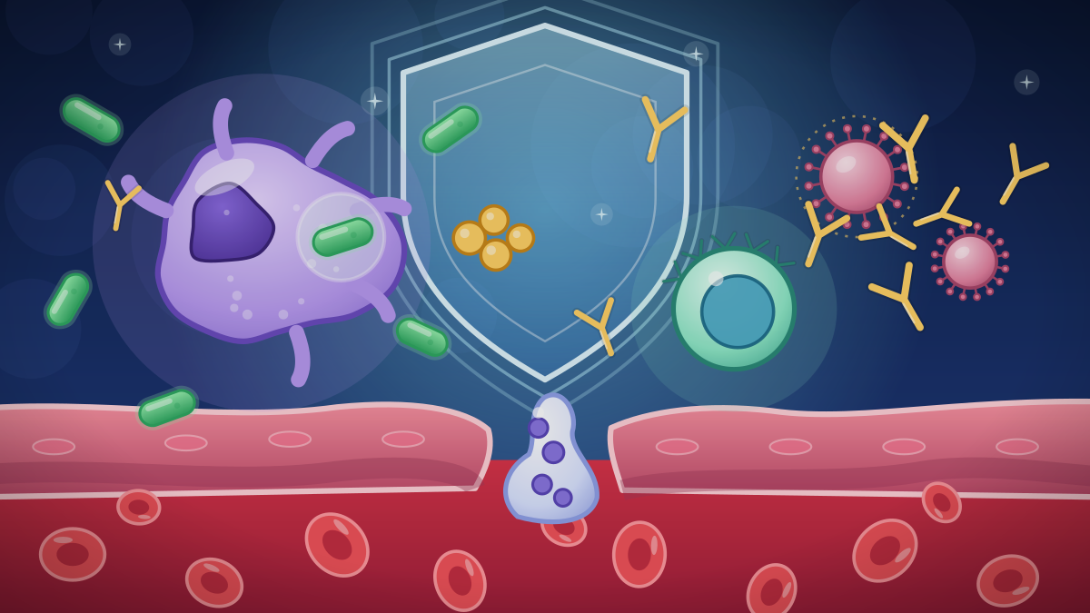
How Does the Immune System Work?
The immune system has a fast, general innate layer and a slower, specific adaptive layer with memory. Barriers, white blood cells, antibodies, organs and what happens when it misfires.
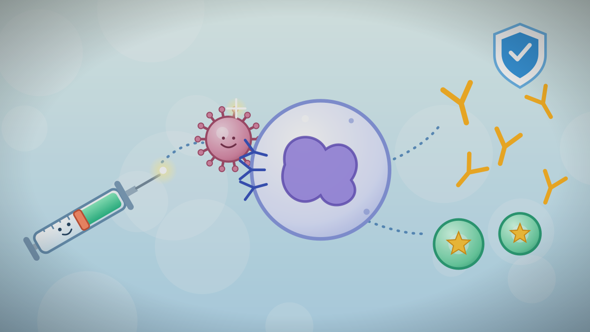
Training the Immune System: How Vaccines Work
Vaccines let the immune system practice against a germ without a real infection. Health agencies describe the antigen, the antibodies and the memory that results.
Why Antibiotics Don't Work on Colds and the Flu
Colds and flu are caused by viruses, and antibiotics do not act on viruses. Public health agencies explain why unnecessary use causes harm.
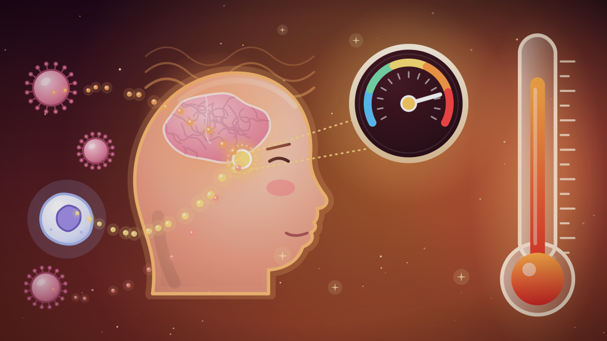
Why Does the Body Get a Fever?
A fever is a raised hypothalamic set point triggered by pyrogens, usually during infection. Health sources describe its mechanism, benefits, how it is graded and when care is advised.
What Are Viruses, and How Do They Differ From Bacteria and Cells?
Viruses are tiny packages of genetic material that cannot run their own metabolism and must hijack host cells to reproduce. Reference texts explain how that sets them apart from bacteria.
References
- OpenStax. Anatomy and Physiology 2e, 21.1 Anatomy of the Lymphatic and Immune Systems. Accessed October 7, 2026.
- OpenStax. Anatomy and Physiology 2e, 21.2 Barrier Defenses and the Innate Immune Response. Accessed October 7, 2026.
- OpenStax. Anatomy and Physiology 2e, 21.3 The Adaptive Immune Response: T Lymphocytes and Their Functional Types. Accessed October 7, 2026.
- OpenStax. Anatomy and Physiology 2e, 21.4 The Adaptive Immune Response: B Lymphocytes and Antibodies. Accessed October 7, 2026.
- OpenStax. Anatomy and Physiology 2e, 20.3 Capillary Exchange. Accessed October 7, 2026.
- CDC. Explaining How Vaccines Work. 2024. Accessed October 7, 2026.
- CDC. About HIV. Accessed October 7, 2026.
- BC Ministry of Education and Child Care. Anatomy and Physiology 12 (curriculum). Accessed October 7, 2026.
These lessons follow the content areas listed in the British Columbia curriculum. They are study material written for this site and are not an official document. The official curriculum is the authority on what each course requires. Lessons are general education, not medical advice.